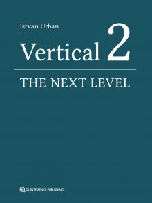
Istvan Urban - Vertical 2: The Next Level of Hard and Soft Tissue Augmentation

Скачивание начинается... Если скачивание не началось автоматически, пожалуйста нажмите на эту ссылку.
Жалоба
Напишите нам, и мы в срочном порядке примем меры.
Описание книги "Vertical 2: The Next Level of Hard and Soft Tissue Augmentation"
Описание и краткое содержание "Vertical 2: The Next Level of Hard and Soft Tissue Augmentation" читать бесплатно онлайн.
A major part of this book comprises full-color, step-by-step images of patient cases. At times, reading it is like watching a surgical video, where the author 'stops the video' to discuss with you, the reader, what he is thinking and doing at that step, what his next step will be, and the reason for it.
Included again are the well-appreciated 'Lessons learned' sections, where the learning objectives are emphasized and further notes given, including ways to further improve the techniques. The section on the mandible is more detailed in this book, with the focus on larger defects and the different surgical steps in native, fibrotic, and scarred tissue types around the mental nerve during flap advancement.
In addition, light is shed on the detail in treating the anterior maxilla, which has not been published previously. It includes treatment options such as the fast track, the safe track, and the technical track of soft tissue reconstruction in conjunction with bone grafting as well as papilla reconstructions after bone regeneration. The section on the posterior maxilla hopes to resolve issues such as the management and complications of combined ridge and sinus grafting, including difficulties such as the lack of buccal, crestal or nasal bony walls of the posterior maxilla before bone grafting.
In this must-have new publication, the procedures are kept simple, repeatable, and biologically sound. The techniques presented are not overcomplicated; they are simple treatment strategies with lower complication rates and more predictability in the final outcome.
Istvan Urban
Vertical 2
Istvan Urban
Vertical 2
THE NEXT LEVEL
OF HARD AND SOFT TISSUE AUGMENTATION
One book, one tree: In support of reforestation worldwide and to address the climate crisis, for every book sold Quintessence Publishing will plant a tree (https://onetreeplanted.org/).
A CIP record for this book is available from the British Library.
ISBN: 978-3-86867-592-4
Quintessenz Verlags-GmbH Quintessence Publishing Co Ltd Ifenpfad 2–4 Grafton Road, New Malden 12107 Berlin Surrey KT3 3AB Germany United Kingdom www.quintessence-publishing.com www.quintessence-publishing.com
Copyright © 2022
Quintessenz Verlags-GmbH
All rights reserved. This book or any part thereof may not be reproduced, stored in a retrieval system, or transmitted in any form or by any means, electronic, mechanical, photocopying, or otherwise, without prior written permission of the publisher.
Editing: Avril du Plessis, Quintessenz Verlags-GmbH, Berlin, Germany
Layout and Production: Janina Kuhn, Quintessenz Verlags-GmbH, Berlin, Germany
Reproduction: Quintessenz Verlags-GmbH, Berlin, Germany
Preface
It has been almost 5 years since the publication of my first book, Vertical and Horizontal Ridge Augmentation: New Perspectives by Quintessence Publishing in 2017. That book has enjoyed great success and has been translated into 12 languages, helping the guided bone regeneration (GBR) technique to be practiced successfully worldwide.
The reader might expect this book to be a second edition. It is not. I had a lot more to share, and this book delves more into the details where the devil lives. I anticipate that you will read this book armed with the knowledge from the first book as regards the anatomy, principles of mandibular surgery, anterior maxillary defect types and their treatment options, and soft tissue reconstruction after bone grafting. It is important that you please review this information from the first book before reading this new one.
Parts of this book are like watching a surgical video with me, where I stop the video at the most important parts (sometimes frame by frame) and discuss with you, the reader, what I am thinking and doing at that step, and what my next step will be. At the same time, I discuss the reason for each of these steps.
In addition, the greatly appreciated ‘Lessons learned’ sections are again included in this book. I consider these sections to be very important, since one can always identify a part of the procedure that one could have done better. These sections also help to emphasize the most important learning objectives of the case.
Please note that one could describe this book as a kind of atlas, a ‘show-and-tell,’ so to speak, where in many places the images, drawings, radiographs, charts, and tables tell the story. For this reason, some chapters contain a minimum of text, and the figures are not always ‘called out’ in the text in a way you may be accustomed to. The idea was to keep things as clear and simple as possible; the figure legends always explain exactly what is going on.
The section on the mandible is more detailed in this book than it was in my first book; also, it focuses on larger defects as well as different surgical steps in native, fibrotic, and scarred tissue types around the mental nerve during flap advancement. The section on the posterior maxilla will hopefully help to solve many issues such as the management of complications of sinus grafting and the lack of buccal, crestal or nasal bony walls of the posterior maxilla before bone grafting.
This book sheds light on the detail in treating the anterior maxilla that has not been published previously. You are finally getting the ‘complete package,’ including treatment options such as the fast track, the safe track or the technical track of soft tissue reconstruction in conjunction with bone grafting. Questions are answered such as: What options do I have when there are multiple implants in regenerated bone and I would like to reconstruct the papilla? The Ice-cube and Iceberg connective tissue graft techniques are the best options, but how do I actually do them, and how do I choose between the two techniques?
I have great expectations for this book, and I really wish that I had had this knowledge two decades ago. I could have had the most perfect cases today. That is what I am expecting for you, dear reader – to make the most perfect cases based on the principles described in this book.
At the same time, as I have said elsewhere, I like to keep procedures simple, repeatable, and biologically sound. The techniques presented here are not overcomplicated – they are simple treatment strategies with lower complication rates and more predictability in the final outcome. Therefore, I would like to welcome you and thank you for reading this book, and remind you of a quote by Leonardo da Vinci: “Simplicity is the ultimate sophistication.”
Some of the cases in this book were not finalized by the time of publication. For additional material and to see the final clinical outcomes of these cases, please scan the QR code on the right or go to the following link:
https://www.quint.link/vertical2mat
Acknowledgments
I would like to thank my family for their love and endless support, and our two sons, Isti and Marci, for their existence, spirit, and positive outlook on life. You make our life complete. As a child (and ever since), my parents never interfered in any of the decisions I made, as they believed in the development of the individual with only minimal guidance. I believe they were right, and I thank them for that.
My teachers, who were my teachers during my training, continue to be my teachers and will remain my teachers. Special thanks to Dr. Henry Takei for his inspiration and unsurpassed qualities as a humanitarian, both as an educator and as a periodontist. Special thanks also to Dr. Jaime Lozada for his belief in me as a student at Loma Linda University and his confidence that I would go on to do vertical ridge augmentation. I would also like to thank Dr. Sascha Jovanovic for introducing me to performing GBR in a biologically sound way. Thanks also to Dr. Joseph Kan, Dr.Perry Klokkevold, Dr. Anna Pogany, Dr. Bela Kovacs, and Dr. Lajos Patonay, and to all my other teachers. Without meeting all of you and being your student, there would be much less to say in this book.
I would like to express my appreciation to Quintessence Publishing, specifically to the management, Horst Wolfgang Haase and Christian Haase.
I would like to express my gratitude to Ms. Krisztina Szample for creating the schematic drawings, and to Denes Doboveczki for assisting in the photography for this book.
I would also like to thank Ms. Jacqueline Kalbach of The Avenues Company for her support in the preparation of my manuscripts, and of this book.
Istvan Urban 2021
Contents
Introduction
Chapter 1 The biology of vertically and horizontally augmented bone
Chapter 2 Scientific evidence of vertical bone augmentation utilizing a titanium-reinforced polytetrafluoroethylene mesh
The extreme vertical defect of the posterior mandible
Chapter 3 Reconstruction of the extreme posterior mandibular defect: surgical principles and anatomical considerations
Chapter 4 Reconstruction of an advanced posterior mandibular defect with scarred tissue
Chapter 5 Reconstruction of an advanced posterior mandibular defect with narrow basal bone
Chapter 6 Reconstruction of an advanced posterior mandibular defect with incomplete periodontal bone levels: the ‘pawn sacrifice’
Chapter 7 Reconstruction of an advanced posterior mandibular defect with the Lasagna technique using low-dose bone morphogenetic protein-
Anterior mandibular vertical augmentation
Chapter 8 Reconstruction of the advanced anterior mandibular defect: surgical principles and anatomical considerations
Chapter 9 Reconstruction of the advanced anterior mandibular defect: considerations for soft tissue reconstruction and preservation of the regenerated bone
Chapter 10 Reconstruction of the advanced anterior mandibular defect: importance of horizontal bone gain
Posterior maxilla
Chapter 11 Long-term results of implants placed in augmented sinuses with minimal and moderate remaining alveolar bone
Chapter 12 Difficulties and complications relating to sinus grafting: hemorrhage and sinus septa
Chapter 13 Difficulties in sinus augmentation and posterior maxillary reconstructions: missing labial sinus wall and ridge deficiencies
Chapter 14 Sinus graft infection and postoperative sinusitis
Chapter 15 The reconstruction of an extreme vertical defect in the posterior maxilla
Anterior maxillary vertical augmentation
Chapter 16 Introduction and clinical treatment guidelines
Chapter 17 Complex reconstruction of an anterior maxillary vertical defect
Anterior maxillary defect
Chapter 18 Extreme defect augmentation in the anterior maxilla
Soft tissue reconstruction in conjunction with bone grafting
Chapter 19 Reconstruction of a natural soft tissue architecture after bone regeneration
Chapter 20 The labial strip gingival graft
Chapter 21 The double strip graft
Chapter 22 Large open-healing connective tissue graft
Reconstruction of the interimplant papilla
Chapter 23 The double connective tissue graft
Chapter 24 The Ice-cube connective tissue graft
Chapter 25 The Iceberg connective tissue graft
Interproximal bone and soft tissue regeneration
Chapter 26 Vertical periodontal regeneration in combination with ridge augmentation
Ultimate esthetics
Chapter 27 Reconstructionof the bone and soft tissue in conjunction with preserving the mucogingival junction
Chapter 28 Complications
Introduction
1
The biology of vertically and horizontally augmented bone
Vertical bone growth is very challenging, both biologically and technically. A recent meta-analysis by Urban et al1 found that, regardless of the technique, about 4.5 mm of vertical bone gain was achieved in the included studies. However, complications occur least frequently when the guided bone regeneration (GBR) technique is used. The results of this study indicate the advantage of using GBR. The technical challenge of GBR is discussed extensively in this book, while the biologic challenge that may play a role in limiting vertical bone gain is discussed in this chapter. Based on the author’s experience, the amount of vertical bone gain is not limited by biology, but more by the clinician’s abilities.
The biologic background of vertical bone gain was investigated by the author in preclinical settings.
The histology of polytetrafluoroethylene (PTFE) membranes in an in vivo setting
The mucogingival tissue is primarily characterized by a moderate vascularized fibrotic reaction that surrounds the PTFE membrane. The membrane is usually internally and externally surrounded by connective tissue that is rich in fibers and poor in cells, with the fibers oriented parallel to the membrane. Membrane pores, if present, show the presence of a highly vascularized matrix of connective tissue and a dense network of collagen fibers that penetrates across the pores to fix on the internal side of the membrane or, in some cases, to the newly formed bone.
The inner layer, which is composed of expanded PTFE (e-PTFE), is always placed against the bone of the lingual and buccal sides. A slight number of macrophages admixed with a few lymphocytes, polymorphonuclear cells, giant cells/osteoclasts, and plasma cells were observed around the membrane. Deep at the lingual side of the ridge, the membrane was often in direct contact with the bone tissue, even showing slight signs of osteointegration (Figs 1-1 to 1-4).
Figs 1-1 and 1-2 Parallel-oriented fibers around the membranes.
Fig 1-3 The membrane shows signs of osseointegration on both sides. Note the excellent biocompatibility of the polytetrafluoroethylene (PTFE) membrane.
Fig 1-4 When compared with titanium, the PTFE-membrane demonstrated similar biocompatibility.
Figs 1-5 and 1-6 Histologic results of vertical augmentation. Note the excellent new bone formation and the well-incorporated xenograft particles.
Bone growth using a xenogenic bone graft
In a preclinical in vivo setting, a chronic vertical defect was treated using a xenograft. After 17 weeks of healing, the following was found: Emerging from the defect bed, the bone growth of a moderate to marked amount showed similar signs of remodeling, resulting in significant vertical ridge augmentation. The bone filler was markedly osteointegrated, showed slight signs of degradation, and demonstrated definite signs of osteoconduction (bone growth on the surface of the granules). The newly formed bone harbored numerous osteoblasts (Figs 1-5 and 1-6).
Подписывайтесь на наши страницы в социальных сетях.
Будьте в курсе последних книжных новинок, комментируйте, обсуждайте. Мы ждём Вас!
Похожие книги на "Vertical 2: The Next Level of Hard and Soft Tissue Augmentation"
Книги похожие на "Vertical 2: The Next Level of Hard and Soft Tissue Augmentation" читать онлайн или скачать бесплатно полные версии.
Мы рекомендуем Вам зарегистрироваться либо войти на сайт под своим именем.
Отзывы о "Istvan Urban - Vertical 2: The Next Level of Hard and Soft Tissue Augmentation"
Отзывы читателей о книге "Vertical 2: The Next Level of Hard and Soft Tissue Augmentation", комментарии и мнения людей о произведении.


